Stress cardiomyopathy - Takotsubo: Difference between revisions
Secretariat (talk | contribs) No edit summary |
No edit summary |
||
| (12 intermediate revisions by 3 users not shown) | |||
| Line 2: | Line 2: | ||
|Title = Takotsubo | |Title = Takotsubo | ||
|CasePresentation = Mimicking myocardial infarction by a stress cardiomyopathy | |CasePresentation = Mimicking myocardial infarction by a stress cardiomyopathy | ||
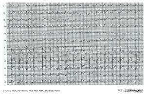
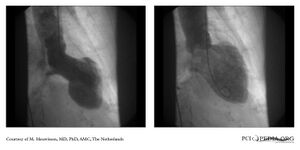
An 81-year-old woman with no cardiac history presented with acute chestpain which radiated to the left arm. The blood pressure was 140/80 mmHg and a heart rate of110/min. Physical examination revealed no abnormalities. The electrocardiogram was compatible with acute anterior myocardial infarction. (A) Immediate coronary angiography showed normal coronary arteries (B and C). A left ventricular (LV) angiogram revealed a Tako-tsubo-like cardiomyopathy, recognized by a hypercontractile base and a bulging out of the LV-apex at systole (D), which normalizes at diastole (E). This typical LV-angiogram resembles a local octopus trap in Japan , where this cardiomyopathy was first described. Although clinical presentation can be quite severe, prognosis is usually good with complete LV recovery. It occurs commonly in post-menopausal woman, usually provoked after extreme emotional stress. In a second interview, the patient told she had a severe emotional experience at a parking lot that morning, after which she developed chestpain. | |||
|Courtesy = Courtesy of M. Meuwissen, MD, PhD, AMC, The Netherlands | |Courtesy = Courtesy of M. Meuwissen, MD, PhD, AMC, The Netherlands | ||
|filepointer1= | |filepointer1=[[Image:MM0100.jpg|300px]] | ||
|file_name1= | |file_name1= | ||
|descriptionfile1= | |descriptionfile1=Figure A: Electrocardiogram | ||
| | |filepointer2=<flash>file=MM0099.swf|quality=best|align=center|width=300|height=300</flash> | ||
| | |file_name2=MM0099 | ||
| | |descriptionfile2=Movie B: Angiography RCA | ||
| | |filepointer3=<flash>file=MM0097.swf|quality=best|align=center|width=300|height=300</flash> | ||
| | |file_name3=MM0097 | ||
| | |descriptionfile3=Movie C: Angiography LCA | ||
| | |filepointer4=<flash>file=MM0098.swf|quality=best|align=center|width=300|height=300</flash> | ||
| | |file_name4=MM0098 | ||
| | |descriptionfile4=Movie D: Left ventricular angiography | ||
| | |filepointer5=[[Image:MM0105.jpg|300px]] | ||
| | |file_name5= | ||
| | |descriptionfile5=Image E: Still frames of left ventricular angiography at end systole and end diastole | ||
}} | }} | ||
Latest revision as of 09:38, 30 August 2009
| Case description: Mimicking myocardial infarction by a stress cardiomyopathy
An 81-year-old woman with no cardiac history presented with acute chestpain which radiated to the left arm. The blood pressure was 140/80 mmHg and a heart rate of110/min. Physical examination revealed no abnormalities. The electrocardiogram was compatible with acute anterior myocardial infarction. (A) Immediate coronary angiography showed normal coronary arteries (B and C). A left ventricular (LV) angiogram revealed a Tako-tsubo-like cardiomyopathy, recognized by a hypercontractile base and a bulging out of the LV-apex at systole (D), which normalizes at diastole (E). This typical LV-angiogram resembles a local octopus trap in Japan , where this cardiomyopathy was first described. Although clinical presentation can be quite severe, prognosis is usually good with complete LV recovery. It occurs commonly in post-menopausal woman, usually provoked after extreme emotional stress. In a second interview, the patient told she had a severe emotional experience at a parking lot that morning, after which she developed chestpain. | |
| Courtesy of: Courtesy of M. Meuwissen, MD, PhD, AMC, The Netherlands | |

|
<flash>file=MM0099.swf |
| Figure A: Electrocardiogram | Movie B: Angiography RCA |
| <flash>file=MM0097.swf | <flash>file=MM0098.swf |
| Movie C: Angiography LCA | Movie D: Left ventricular angiography |

|
|
| Image E: Still frames of left ventricular angiography at end systole and end diastole | |